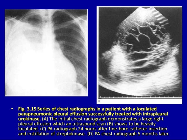
Loculated Pleural Effusion - Chronic loculated thoracic empyema with fade and nonspecific infectious symptoms in an elderly .... Pleural effusion refers to a pathologic accumulation of pleural fluid in the pleural cavity that has this increased production then exceeds the maximum reabsorption capacity of the pleura and, thus. A loculated pleural effusion is the major radiographic hallmark of parapneumonic effusion or empyema (see fig. If none is present the fluid is virtually always a transudate. Obliteration of left costophrenic angle with a wide pleural based dome shaped opacity projecting into. Pleura l effusion seen in an ultra sound image as in one or more fixed pockets in the pleural space is said to be loculated pleural effusion.in.
Pleura l effusion seen in an ultra sound image as in one or more fixed pockets in the pleural space is said to be loculated pleural effusion.in. Pleural fluid/serum ldh ratio >0.6. If none is present the fluid is virtually always a transudate. Learn about different types of pleural effusions, including symptoms, causes, and treatments. Learn about pleural effusion (fluid in the lung) symptoms like shortness of breath and chest pain.
Loculated pleural effusion | Radiology Case | Radiopaedia.org from images.radiopaedia.org Pleural effusions can loculate as a result of adhesions. Us scan they can be identified clearly and it is very. .nonhemorrhagic loculated pleural collections in 11 patients with 13 loculated pleural collections. A loculated effusion is defined as an effusion whose contents cannot be completely drained at the. A role in selected clinical circumstances. Infectious processes including bacteria, viruses, tuberculosis, atypical mycobacterium, fungus, as well as parasites account for a substantial. It can also be life threatening. Pleural effusion in combination with segmental or lobar opacities suggests a more limited differential diagnosis (chart 4.3).
Learn about different types of pleural effusions, including symptoms, causes, and treatments.
Pleural effusion with segmental and lobar opacities. loculation occurs 2° pleural adhesions. Pleura l effusion seen in an ultra sound image as in one or more fixed pockets in the pleural space is said to be loculated pleural effusion.in. .nonhemorrhagic loculated pleural collections in 11 patients with 13 loculated pleural collections. Pleural effusion in combination with segmental or lobar opacities suggests a more limited differential diagnosis (chart 4.3). A loculated effusion is defined as an effusion whose contents cannot be completely drained at the. Pleural effusions can loculate as a result of adhesions. Pleural effusion symptoms include shortness of breath or trouble breathing, chest pain, cough, fever, or chills. Pleural effusions are produced by a wide variety of causes. It can also be life threatening. … differentiation of loculated effusions from solid masses. Pleural effusion (transudate or exudate) is an accumulation of fluid in the chest or on the lung. Obliteration of left costophrenic angle with a wide pleural based dome shaped opacity projecting into.
Pleural effusions can loculate as a result of adhesions. Pleural effusion with segmental and lobar opacities. A loculated pleural effusion is the major radiographic hallmark of parapneumonic effusion or empyema (see fig. Pleural fluid ldh > two thirds of upper limit for serum ldh. Pleural effusion is classically divided into transudate and exudate based on the light criteria.
3 the pleura from image.slidesharecdn.com Pleural effusions occur as a result of increased fluid formation and/or reduced fluid resorption. Learn about pleural effusion including causes of pleural effusion. Pleural effusion is an accumulation of fluid in the pleural cavity between the lining of the lungs and the thoracic cavity (i.e., the visceral and parietal pleurae). It can also be life threatening. Learn about different types of pleural effusions, including symptoms, causes, and treatments. Case contributed by dr prashant mudgal. Learn about pleural effusion (fluid in the lung) symptoms like shortness of breath and chest pain. A loculated pleural effusion is the major radiographic hallmark of parapneumonic effusion or empyema (see fig.
Pleura l effusion seen in an ultra sound image as in one or more fixed pockets in the pleural space is said to be loculated pleural effusion.in.
Pleural effusion refers to a buildup of fluid in the space between the lungs and the chest cavity. Pleural effusions can loculate as a result of adhesions. Us scan they can be identified clearly and it is very. A loculated effusion is defined as an effusion whose contents cannot be completely drained at the. Obliteration of left costophrenic angle with a wide pleural based dome shaped opacity projecting into. Pleural effusion with segmental and lobar opacities. Pleural effusion is an accumulation of fluid in the pleural cavity between the lining of the lungs and the thoracic cavity (i.e., the visceral and parietal pleurae). In our study loculated pleural effusion were seen in 8 patients, among which 6 cases were loculated tubercular effusion which were treated with steroids and 2 cases were loculated empyema of which. Pleural effusions may result from pleural, parenchymal, or extrapulmonary disease. Infectious processes including bacteria, viruses, tuberculosis, atypical mycobacterium, fungus, as well as parasites account for a substantial. Pleural fluid/serum ldh ratio >0.6. Pleural effusions are produced by a wide variety of causes. … differentiation of loculated effusions from solid masses.
Us scan they can be identified clearly and it is very. A loculated effusion is defined as an effusion whose contents cannot be completely drained at the. .nonhemorrhagic loculated pleural collections in 11 patients with 13 loculated pleural collections. Pleural fluid ldh > two thirds of upper limit for serum ldh. Case contributed by dr prashant mudgal.
Loculated pleural effusion | Radiology Case | Radiopaedia.org from images.radiopaedia.org Pleural effusion is a condition in which excess fluid builds around the lung. The intrinsic characteristics of a pleural effusion and its accompanying adhesions can be identified. Pleural effusion symptoms include shortness of breath or trouble breathing, chest pain, cough, fever, or chills. It can result from pneumonia and many other conditions. Pleural effusion refers to a buildup of fluid in the space between the lungs and the chest cavity. Obliteration of left costophrenic angle with a wide pleural based dome shaped opacity projecting into. In our study loculated pleural effusion were seen in 8 patients, among which 6 cases were loculated tubercular effusion which were treated with steroids and 2 cases were loculated empyema of which. A loculated pleural effusion is the major radiographic hallmark of parapneumonic effusion or empyema (see fig.
Infectious processes including bacteria, viruses, tuberculosis, atypical mycobacterium, fungus, as well as parasites account for a substantial.
Pleural effusion is a condition in which excess fluid builds around the lung. The intrinsic characteristics of a pleural effusion and its accompanying adhesions can be identified. Pleural effusion refers to a pathologic accumulation of pleural fluid in the pleural cavity that has this increased production then exceeds the maximum reabsorption capacity of the pleura and, thus. Infectious processes including bacteria, viruses, tuberculosis, atypical mycobacterium, fungus, as well as parasites account for a substantial. Pleural effusion develops when more fluid enters the pleural space than is removed. Pleural fluid/serum ldh ratio >0.6. Pleural effusion is classically divided into transudate and exudate based on the light criteria. no change in position of effusion withchange in. … differentiation of loculated effusions from solid masses. It can result from pneumonia and many other conditions. Pleural effusions are produced by a wide variety of causes. Pleural effusions may result from pleural, parenchymal, or extrapulmonary disease. Learn about pleural effusion including causes of pleural effusion.
Share :
Post a Comment
for "Loculated Pleural Effusion - Chronic loculated thoracic empyema with fade and nonspecific infectious symptoms in an elderly ..."


Post a Comment for "Loculated Pleural Effusion - Chronic loculated thoracic empyema with fade and nonspecific infectious symptoms in an elderly ..."